This acclaimed book presents neuroanatomy through over 100 clinical cases and high-quality radiologic images, offering an interactive, engaging approach ideal for medical students and neuroscientists. Available as a free PDF, it effectively integrates anatomy with real-world scenarios, making complex concepts accessible and practical.
Overview of the Book and Its Approach
Neuroanatomy Through Clinical Cases offers a pioneering, interactive approach to teaching neuroanatomy. The book integrates anatomical and clinical content through over 100 real clinical cases and high-quality radiologic images. This method bridges theory and practice, making neuroanatomy engaging and accessible. The third edition is fully updated, incorporating the latest advances in the field. It is designed for medical students and neuroscientists, providing a comprehensive yet practical understanding of neuroanatomy.
Importance of Clinical Cases in Neuroanatomy Education
Clinical cases are essential in neuroanatomy education as they bridge theory and practice. By presenting real-life scenarios, they help students understand the practical relevance of neuroanatomy. Over 100 clinical cases in the book, supported by radiologic images, create an engaging and interactive learning experience. This approach enhances retention and application of knowledge, making neuroanatomy more accessible and meaningful for medical students and neuroscientists;

Structure and Organization of the Book
The book is logically structured, starting with basic neuroanatomy and progressing to advanced topics. It includes clinical cases, radiologic images, and interactive elements, making it comprehensive.
Key Features of the Third Edition
The third edition includes over 100 clinical cases, high-quality radiologic images, and interactive pedagogy. It features focused questions, case discussions, and updated content reflecting the latest advances in neuroanatomy. The book is available as a free PDF, making it accessible for medical students and neuroscientists. Its structured approach integrates anatomy with clinical scenarios, enhancing learning and retention. The edition also incorporates feedback from previous versions, ensuring improved clarity and relevance.
Integration of Anatomical and Clinical Content
The book seamlessly integrates neuroanatomical concepts with real clinical scenarios, creating a comprehensive learning experience. By linking structural details to patient cases, it helps students understand how neuroanatomy applies to diagnosis and treatment. This approach fosters a deeper appreciation of the subject, making it both practical and engaging for medical students and neuroscientists. The integration enhances the ability to correlate anatomical knowledge with clinical presentations effectively.
Clinical Case Studies and Radiologic Images
The book features over 100 clinical cases with high-quality radiologic images, providing a comprehensive and visual approach to understanding neuroanatomy in a real-world context.
Overview of the 100+ Clinical Cases Presented
The book includes over 100 clinical cases, each accompanied by high-quality radiologic images, covering a wide range of neurological conditions. These cases are designed to illustrate key neuroanatomical concepts through real-life examples, making the subject more engaging and accessible for learners. Each case integrates anatomical and clinical information, providing a comprehensive understanding of brain function and dysfunction. This approach helps students and professionals connect theoretical knowledge with practical patient scenarios, enhancing their diagnostic and clinical reasoning skills.
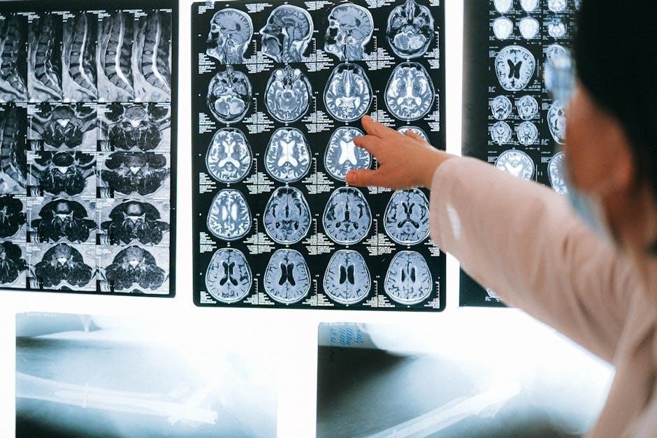
Role of High-Quality Radiologic Images in Learning
High-quality radiologic images play a crucial role in enhancing neuroanatomy education by providing visual confirmation of anatomical structures. These images, often MRI and CT scans, allow learners to correlate theoretical knowledge with real-life brain anatomy and pathology. The clarity and detail of these images help students identify key structures, understand their spatial relationships, and grasp the clinical relevance of neuroanatomical concepts, making learning more effective and engaging.

The Neurological Exam as a Teaching Tool
The neurological exam serves as a dynamic teaching tool, linking clinical assessment findings to specific neuroanatomical structures and their functions, making learning interactive and clinically relevant.
Basic Definitions and Concepts in Neuroanatomy
Neuroanatomy begins with understanding the brain’s structural components, such as neurons, glial cells, and the blood-brain barrier. Key concepts include the organization of the nervous system into central and peripheral divisions. These foundational elements are essential for grasping how neuroanatomical structures relate to clinical cases, enabling learners to connect basic science with real-world neurological disorders and their diagnoses.
How the Neurological Exam Illustrates Neuroanatomy
The neurological exam serves as a practical tool, bridging neuroanatomy and clinical practice. By assessing functions like motor strength, reflexes, and sensory perception, it illuminates the brain’s structural and functional organization. Each examination finding correlates with specific neuroanatomical pathways, enabling precise localization of lesions and enhancing diagnostic accuracy. This integration transforms abstract concepts into actionable insights, making neuroanatomy accessible and relevant for clinicians and students alike.

Interactive Pedagogy and Learning Techniques
The book employs case-based learning, focused questions, and interactive discussions to engage students, fostering active learning and the practical application of neuroanatomy in clinical contexts effectively.
Engaging Students Through Interactive Learning
The book uses interactive pedagogy, including case-based learning and focused questions, to actively engage students. Over 100 clinical cases and high-quality radiologic images are presented, making neuroanatomy relatable and practical. This approach encourages students to think critically and apply knowledge in real-world scenarios, enhancing understanding and retention. The interactive format makes learning dynamic and participatory, helping students grasp complex concepts through hands-on problem-solving and discussion. The free PDF availability further increases accessibility for a broader audience.
Use of Focused Questions and Case Discussions
The book employs focused questions and case discussions to deepen understanding. Each clinical case is accompanied by targeted questions that guide students through diagnostic reasoning. This approach encourages active learning, helping students apply neuroanatomical knowledge to real patient scenarios. The discussions facilitate critical thinking and collaboration, fostering a deeper grasp of complex concepts. This method ensures students are well-prepared for clinical practice, bridging the gap between theory and application effectively.

Target Audience and Educational Use
Primarily designed for medical students and neuroscientists, this book is ideal for neuroanatomy or neuroscience courses. It integrates clinical cases and radiologic images to enhance learning, making it a valuable resource for educational purposes and professional development.
Primary Audience: Medical Students and Neuroscientists
This book is specifically tailored for medical students and neuroscientists, particularly those in their early years of study. It serves as an essential resource for courses in neuroanatomy or neuroscience, offering a unique blend of clinical cases and radiologic images. The interactive approach and real-world applications make it ideal for learners seeking to bridge theoretical knowledge with practical understanding. Its availability as a free PDF further enhances accessibility for students and professionals alike.
Suggested Course Integration and Study Tips
This book is designed for medical students and neuroscientists, offering a practical approach to neuroanatomy through clinical cases. Instructors can integrate its chapters into neuroscience or neuroanatomy courses, supplementing lectures with case discussions. Students are encouraged to review radiologic images and correlate them with clinical presentations. Focused questions and interactive pedagogy enhance understanding, while the free PDF format allows easy access for study and review.
Author and Contributions
Hal Blumenfeld, a renowned neuroanatomy expert and Yale professor, authored this seminal work, integrating clinical cases and radiologic images to enhance learning. His collaborative approach and contributions have made the book a cornerstone in neuroanatomy education.
Hal Blumenfeld and His Expertise in Neuroanatomy
Hal Blumenfeld, a distinguished Yale professor and epilepsy researcher, brings unparalleled expertise to neuroanatomy education. His groundbreaking approach combines clinical cases and radiologic images, making complex concepts accessible. With years of experience in both research and teaching, Blumenfeld has developed a unique ability to engage students through real-world applications, ensuring a deeper understanding of neuroanatomy.
Acknowledgments and Collaborations in the Book
The book acknowledges contributions from neurologists, neurosurgeons, and radiologists who provided clinical cases and images. Collaborations with experts ensure content accuracy and relevance, enriching the learning experience. Blumenfeld expresses gratitude to colleagues and students, highlighting teamwork’s role in creating a comprehensive resource for neuroanatomy education.

Availability and Access
The book is widely available for free download as a PDF from platforms like Medicalstudyzone.com and uDocz, ensuring easy access to its comprehensive content.
Downloading the PDF for Free
The PDF version of “Neuroanatomy Through Clinical Cases” is readily available for free download on various platforms such as Medicalstudyzone.com and uDocz. These websites provide direct links to the full-text PDF, allowing users to access the book instantly. The third edition is also accessible, ensuring that students and professionals can benefit from the latest updates and advancements in the field without any cost barriers. This accessibility makes the resource widely popular among medical students and neuroscientists seeking a comprehensive understanding of neuroanatomy through real-world clinical scenarios.
Edition Updates and Latest Advances in Neuroanatomy
The third edition of “Neuroanatomy Through Clinical Cases” incorporates the latest advances in neuroanatomy, featuring updated clinical cases and high-quality radiologic images. It includes new insights into neurological disorders and diagnostic techniques, ensuring students and professionals stay current with modern developments. The revised content bridges educational concepts with real-world applications, enhancing the learning experience and clinical relevance for medical students and neuroscientists alike.
Impact on Medical Education
Blumenfeld’s interactive approach significantly enhances medical education by making neuroanatomy engaging and accessible through real-life clinical cases and high-quality images, fostering deeper understanding and practical application for students.
Student Feedback and Perceived Benefits
Students highly praise the book for its clarity and practical approach, noting improved understanding of neuroanatomy through real-life clinical cases and high-quality images. The interactive format and focused questions enhance learning, making complex concepts more accessible. Many appreciate how the integration of anatomy with clinical scenarios bridges theory and practice, aiding in exam preparation and patient care. This approach is widely regarded as a valuable resource for medical education.
Blumenfeld’s Approach to Making Neuroanatomy Engaging
Hal Blumenfeld’s innovative approach combines over 100 clinical cases, high-quality radiologic images, and interactive pedagogy to engage students. By focusing on real patient scenarios, he bridges anatomy with clinical practice, making neuroanatomy relatable and practical. This method not only enhances understanding but also fosters critical thinking, preparing students for both exams and future patient care with a deeper appreciation of neuroanatomy’s clinical relevance.
Neuroanatomy Through Clinical Cases offers a groundbreaking approach, blending clinical scenarios with anatomical insights. Its free PDF availability makes it a valuable resource for learners seeking an engaging, practical understanding of neuroanatomy.
Final Thoughts on the Book’s Value
Neuroanatomy Through Clinical Cases is a transformative resource that bridges neuroanatomy and clinical practice through engaging case studies and high-quality images. Its interactive approach and free PDF availability make it accessible to all learners. By integrating real-world scenarios with anatomical insights, this book revolutionizes neuroanatomy education, proving invaluable for medical students, neuroscientists, and professionals seeking a deeper understanding of the brain and its functions.
Encouragement to Explore the Full Text
Exploring the full text of Neuroanatomy Through Clinical Cases offers unparalleled insights into the brain’s structure and function. With over 100 clinical cases and high-quality radiologic images, this resource provides a unique, interactive learning experience. Ideal for medical students and neuroscientists, it bridges theory and practice, making neuroanatomy accessible and engaging. Download the free PDF to delve into its comprehensive content and enhance your understanding of the nervous system through real-world applications.